this month · 11 submission slots remaining — reviewed in order of receipt
Your iris is
the most detailed map
of yourself that exists.
Upload a photo of your iris. Receive a 20+ page structural analysis of your constitutional type, constitutional zones, and psycho-emotional history.
Most people spend a lifetime making decisions about their health, energy, and emotions — without ever reading the structural map they were born with.

protocol
01 — CAPTURE
Photograph your iris
Smartphone camera in natural daylight. No equipment, no appointment, no specialist required. Follow the illustrated guide on the analysis page for lighting, distance, and focus.
02 — SUBMIT
Upload and position
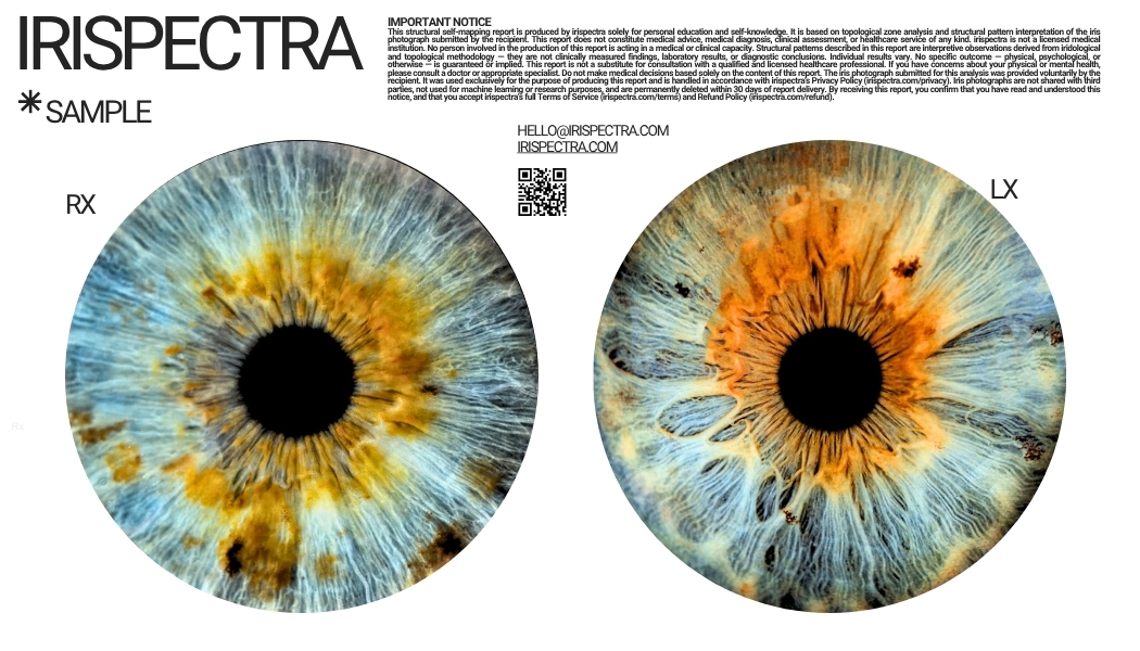
Submit both iris photographs through the secure intake. Use the built-in tools to align each image precisely within the circular mapping guide. Right eye (RX) and left eye (LX) both required.
03 — RECEIVE
Your map is delivered
A structured PDF — constitutional architecture, zone patterns, psycho-emotional timeline, and individualised protocols. Delivered within 3 hours of payment. Payment link sent only after image review.
→ No payment until your images have been reviewed
scientific basis
The eye as a
systemic biomarker window.
The retina is the only location in the human body where microcirculation is directly observable non-invasively. Retinal vascular structures reflect cardiovascular, metabolic, and neurological status with measurable precision.
The iris, connected to the body via the autonomic nervous system, encodes structural and pigmentation patterns corresponding to constitutional type, constitutional vitality, and psycho-emotional history.
detection accuracy by condition — peer-reviewed studies
report contents
A precision map
of your constitutional self.
Every irispectra report integrates structural pattern analysis, Rayid constitutional methodology, and topological zone mapping. The result is a structured document — not a generic wellness summary, but a map specific to the structural patterns visible in your iris.
The psycho-emotional timeline is unique to irispectra. Zone markings are cross-referenced to developmental age windows — surfacing structural patterns that correspond to specific periods in your personal history. Clients consistently describe this section as the most precise thing they have ever read about themselves.
sample findings
Choose your
depth.
Each mapping begins with iris submission. Images are reviewed before any payment is requested — you only pay once your photographs have been confirmed suitable for analysis.
Weekly capacity is limited. Submissions are reviewed in order of receipt.
Precision Report
For a first structural read of yourself.
Complete bilateral structural analysis. A full self-mapping report delivered as a structured PDF.
one-time · 3 hour delivery
- Both eyes mapped independently (RX + LX)
- 47+ zone topological analysis
- Constitutional architecture profile
- Psycho-emotional developmental timeline
- Constitutional zone structural mapping
- Individualised protocols and guidance
- Downloadable structured PDF report
no payment until images are reviewed
Precision Monthly
For those actively refining their wellness protocols.
Full report plus monthly structural re-assessment and direct practitioner access.
per month · cancel anytime
- Everything in Precision Report
- Monthly structural re-assessment
- Protocol updates over time
- Direct access via email and WhatsApp
- Progress tracking across sessions
- Priority report turnaround
- Lifestyle and protocol guidance
select monthly in the submission form
Precision Deep
For deep personal work with guided interpretation.
Full structural report plus a live 60-minute guided session and three-month follow-up protocol.
single engagement
- Full Precision Report included
- 60-minute live session via video
- Session recording provided
- Three-month follow-up protocol
- Priority scheduling
- Direct practitioner access throughout
limited availability · by application
No appointment. No waiting room. No blood draw. Delivered in 3 hours.
order process
01
Submit your iris photos
02
Images reviewed
quality confirmed before payment
03
Payment link sent
only after review confirmation
04
Precision report delivered
structured PDF, 3 hours
feedback
"The psycho-emotional timeline identified a developmental period I had never consciously connected to my current patterns. The structural specificity was unlike anything I have encountered."
Dr. Elif K.
Integrative Wellness, Istanbul
"I now use irispectra as a preliminary intake tool. Constitutional mapping gives patients an immediate structural self-awareness that shortens the entire therapeutic arc."
M. Bergmann
Naturopath, Berlin
"I had done genetic testing, blood panels, and extensive functional work. irispectra surfaced structural patterns none of it had named. The precision was unexpected."
Sarah L.
Health Optimisation, London
Common
questions.
hello@irispectra.com
Important Disclaimer
irispectra produces structural self-mapping reports for personal education and self-knowledge. Our reports are not advice, diagnosis, treatment, or healthcare services of any kind. irispectra is not a licensed institution. Results are interpretive, based on structural pattern and topological methodology, and are intended solely for informational and educational purposes. We do not claim to diagnose, treat, cure, or prevent any condition. Structural patterns are interpreted, not measured. Always consult a qualified healthcare professional for concerns. By submitting your photographs, you confirm that you have read and understood this notice.
No payment until images are reviewed
Upload your iris photos →